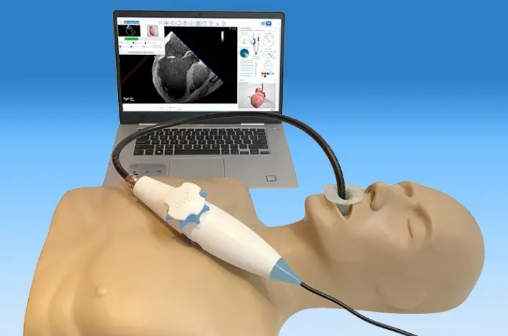
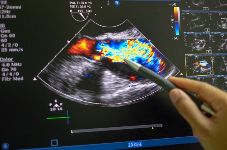
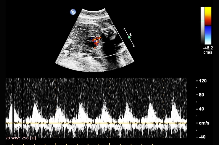
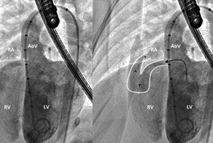
Real-time heart imaging during surgery, guiding surgeons with immediate feedback on cardiac function, valve repairs, and structural corrections.
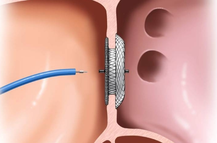
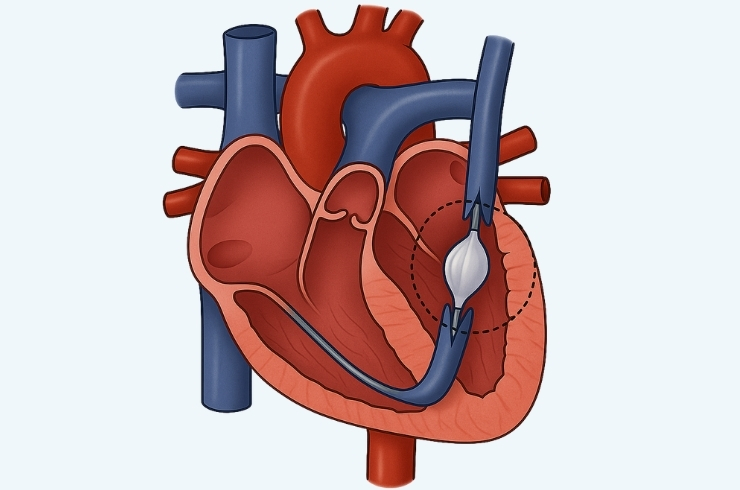
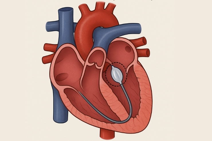
Catheter-based procedure where a balloon is carefully inflated to widen narrowed heart valves or vessels, significantly improving blood flow and function.